
PRODUCT
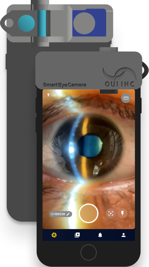
Smart Eye Camera
Ophthalmic Exams Via Your Smartphone
Anytime, Anywhere, and by Anyone


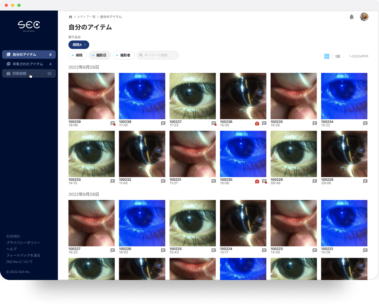
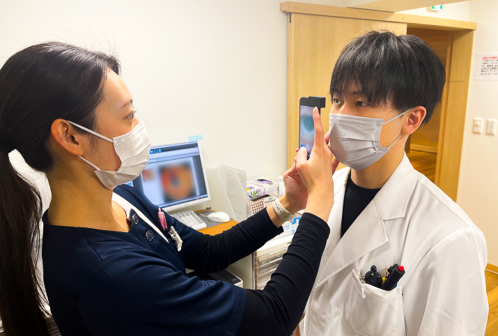
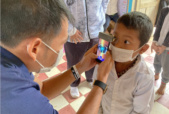
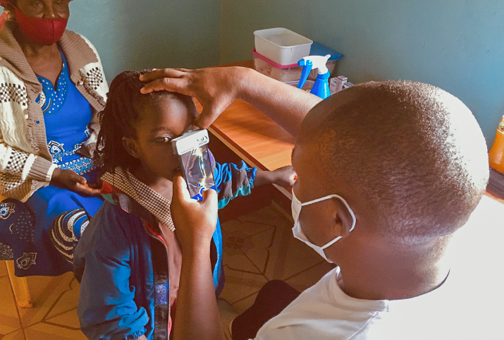
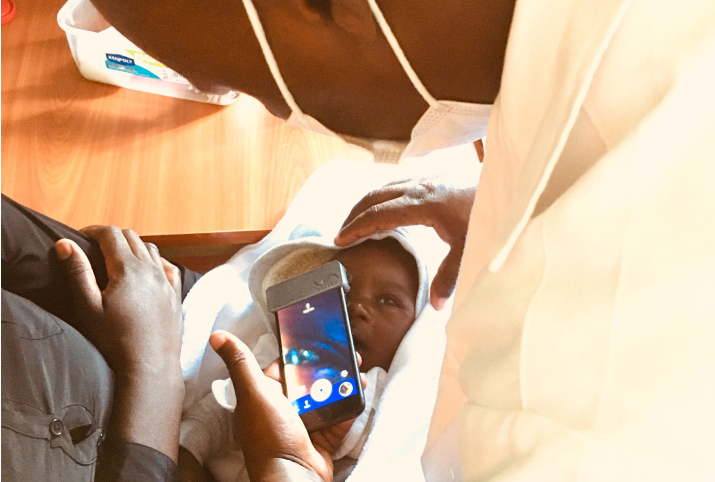
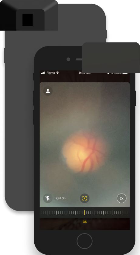
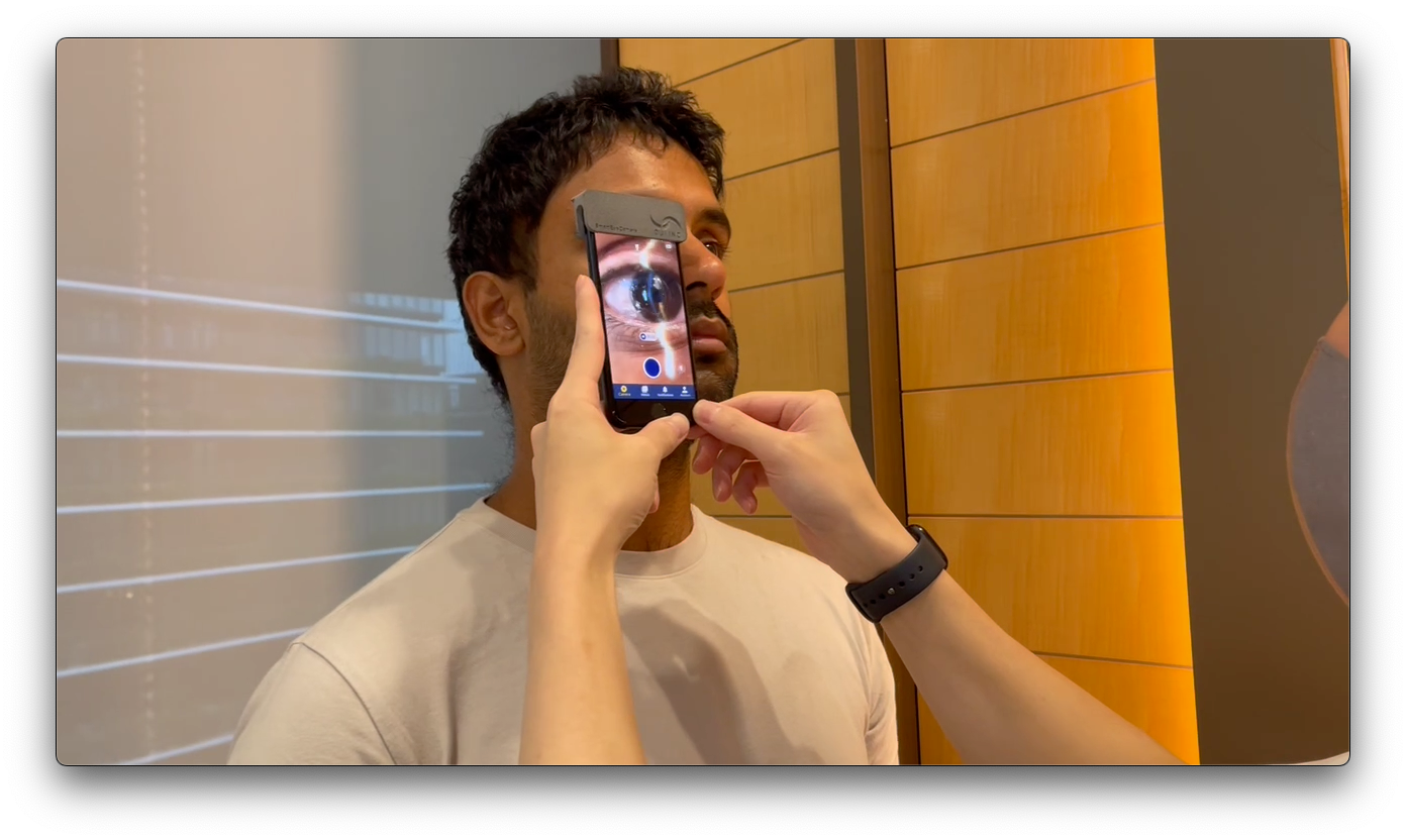
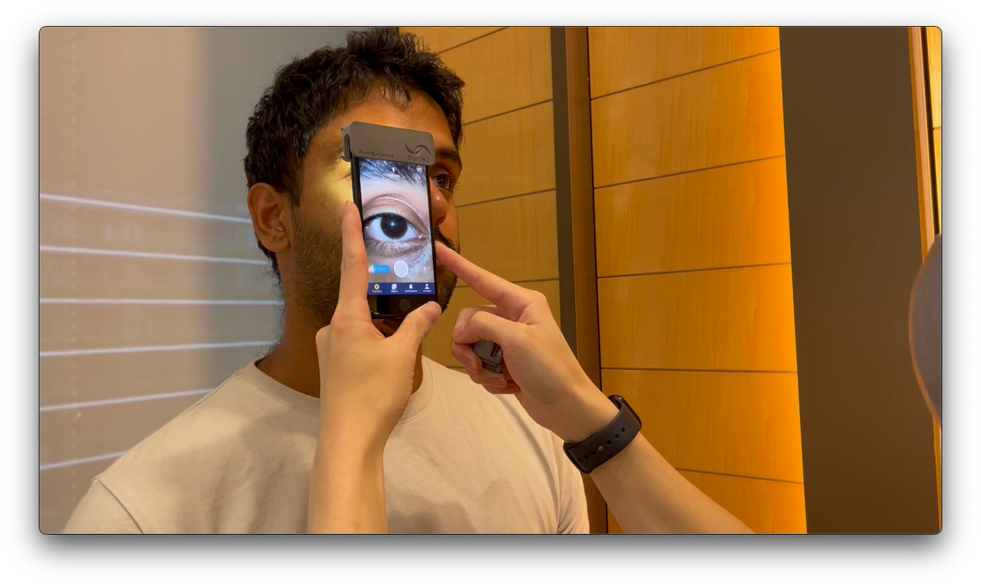
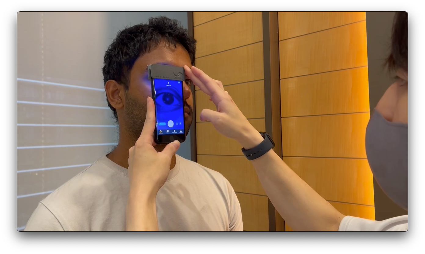
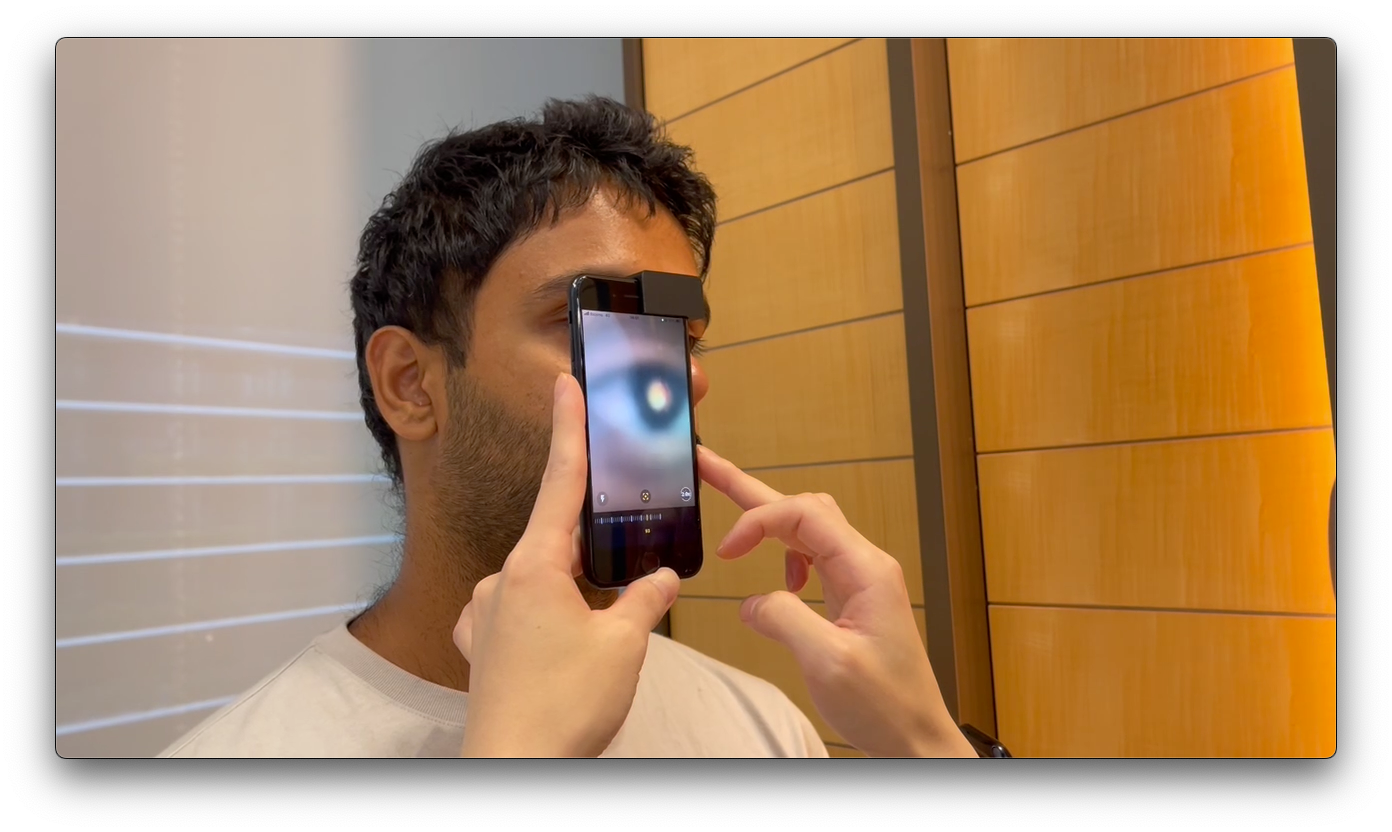
The Smart Eye Camera, SEC, is an ophthalmic medical device, invented by Dr. Eisuke, a practicing ophthalmologist in Japan, to solve his own diagnostic problems in Japan and developing nations. The SEC is a smartphone attachment which requires no additional tools. It allows you to observe the eyelids, cornea, conjunctiva, anterior chamber, iris, lens, and vitreous body in the same manner as a conventional slit lamp microscope. This further enables you to diagnose various diseases, such as cataracts. Additionally, using a dedicated application for image filing, remote ophthalmic diagnosis and the consequential treatment are also possible. The creation of the SEC has made it possible for anyone to perform eye examinations at any time, regardless of location.
The global blindness prevalence is 44 million, and it is projected to roughly triple to 120 million in the next 30 years. More than half of the cases are caused by cataracts, which is treatable with first a proper diagnosis. Unfortunately due to lack of access to resources, this treatable disease goes untreated very often. This is where the SEC comes in - providing accessible and affordable means of screening cataract and other diseases.
This way, OUI Inc.'s portable and convenient device, the SEC, can play a major role in reducing worldwide blindness to a huge extent.
information Publication Information
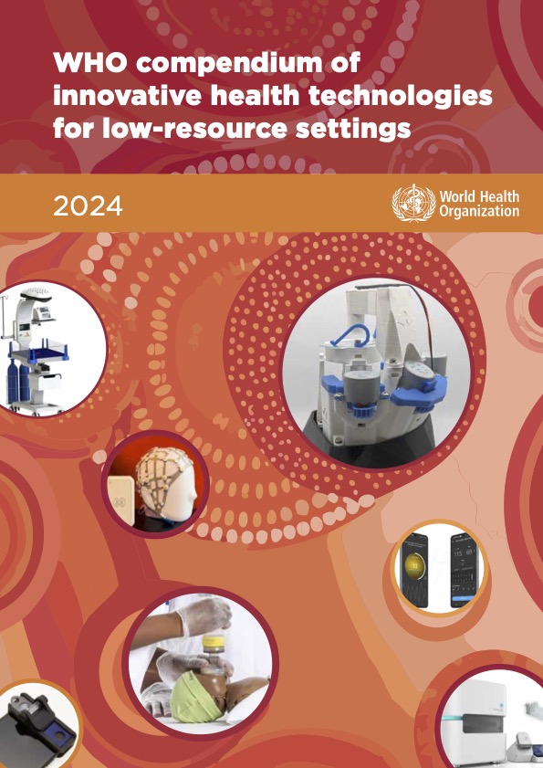
The SEC is the only medical device listed in Japan in the WHO compendium of innovative health technologies for low-resource settings 2024.
The "WHO Compendium of Innovative Health Technologies for Low-Resource Settings 2024" is a catalog of medical devices, recommended for developing countries. It aims to distribute innovative health technologies to a wider patient population, by providing reliable evaluations and relevant information.
 slit lamp
Just by attaching the attachment to your smartphone, you can perform an anterior eye examination in the same way as through a conventional slit lamp microscope! You also don't have to worry about an external light source or replacing a battery, as those are obtained from the smartphone itself. The set up fits in the pocket of your white coat.
Further on, as shown below, we have a growing list of studies using the SEC, serving as evidence for safety and comparability in performance to a conventional slit lamp microscope.
Lastly, under the Gray Zone Elimination system, medical imaging, remote diagnosis, and treatment are possible.
- Examination of the anterior segment of the eye
- Telemedicine is possible
- Insurance points (Japan) for healthcare professionals can be gained
 ophthalmoscope
Just by attaching the attachment to your smartphone, you can examine the fundus of an eye! The optic disk and the posterior pole are visible WITHOUT dilation. You also don't have to worry about an external light source or replacing a battery, as those are obtained from the smartphone itself. The set up fits in the pocket of your white coat.
Download Catalog- Observation of the fundus
- Examination of the optic disk and the posterior pole W/O mydriasis
- Insurance points (Japan) for healthcare professionals can be gained
capabilities The Smart Eye Camera Makes It Possible To
-


Eye Diagnosis and Treatment via Your Smartphone
Through attaching the attachment to your smartphone, you can perform ophthalmic diagnosis. There is no need for an external light source or an external battery, fitting in your pocket! Remote medical diagnosis and treatment is possible via the internet!
-

Diagnose Ophthalmic Diseases Easily
The slit lamp model is suitable for observing the anterior segment of the eye, including the eyelids, cornea, conjunctiva, anterior chamber, iris, and crystalline lens.
It is useful for many ophthalmic diseases such as cataracts, dry eyes, allergic conjunctival diseases, and evaluating the anterior chamber depth. -

3D Printed
All SEC models are manufactured using high-precision 3D printers.
It doesn't require a battery and has minimally moving parts, so it's durable and portable.
-
Slit Light
By setting the SEC to the slit light mode, it is possible to diagnose cataracts, estimate the depths of the anterior chamber, as well as diagnose narrow angle glaucoma attacks.
-
White Diffuse Light
By removing the slit and the blue filter, it is possible to diagnose allergic conjunctival diseases and to fit soft contact lenses, as well.
-
Blue Diffuse Light
By installing the blue filter, it is possible to diagnose diseases such as the dry eye disease and herpetic keratitis, and also to fit ortho-k lenses.
-
Mydriasis
The optic disc and posterior pole can be observed without mydriasis, making it possible to observe risk factors for glaucoma such as the enlargement of the cup-to-disc ratio (cupping)..
cases Filming scenery
-
Slit light
-
White diffuse light
-
Blue diffuse light
-
Mydriasis
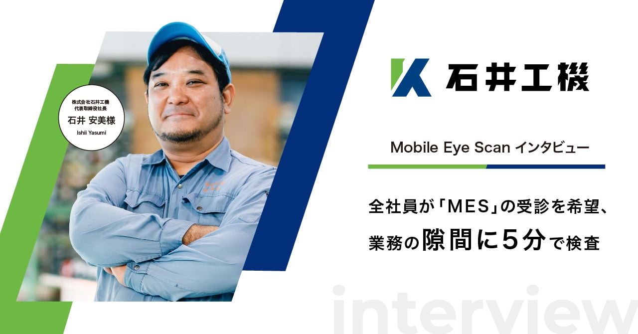
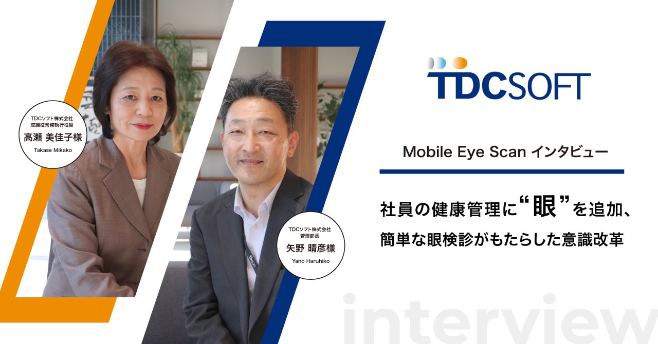
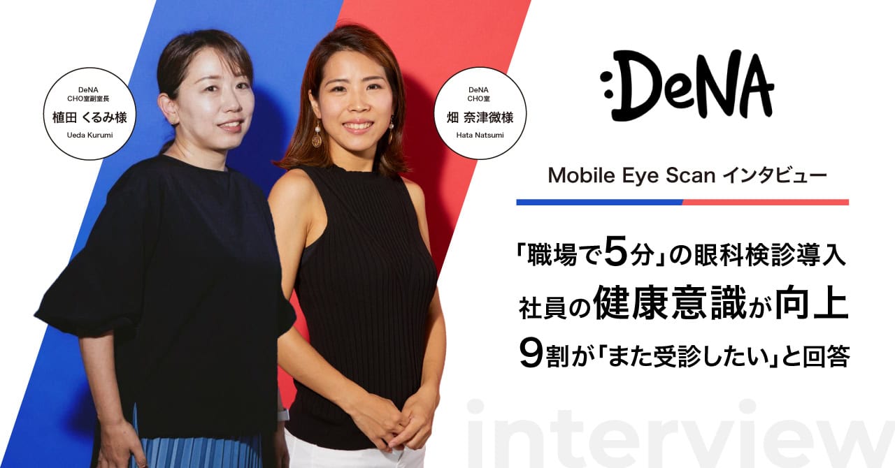
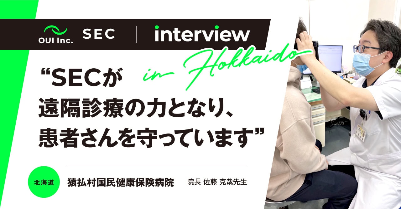
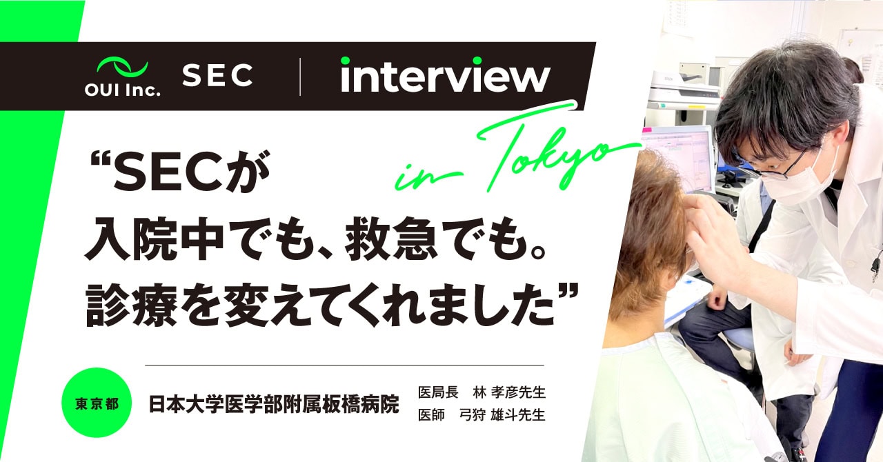
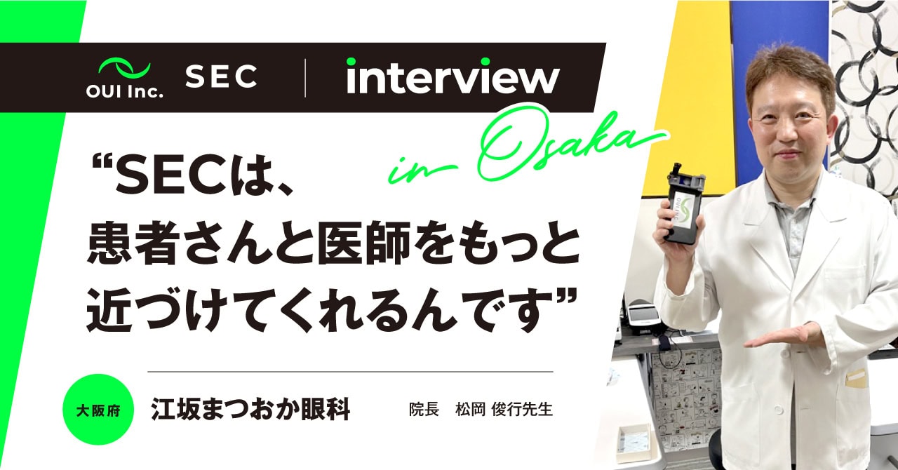
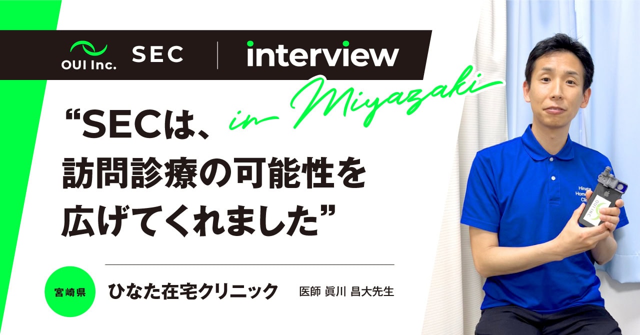
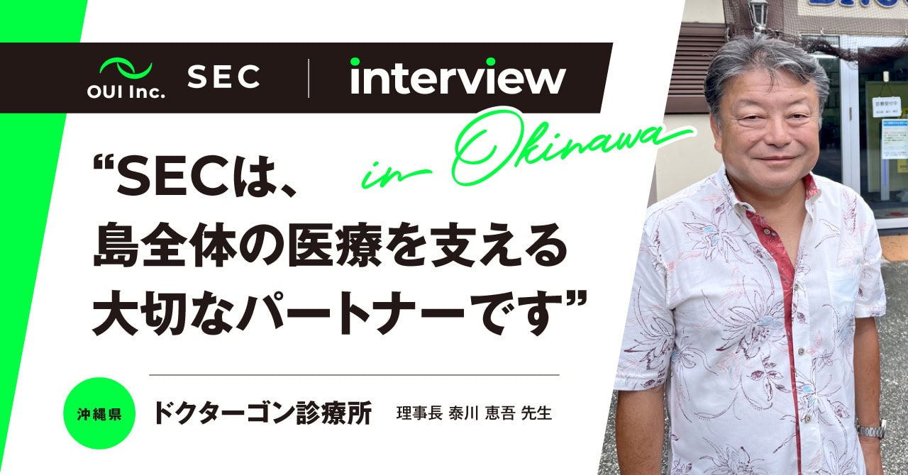
interview Interview Case Studies
youtube YouTube Case Studies
-
slit lamp
Epidemic Keratoconjunctivitis
-
slit lamp
Dry Eye Disease
-
slit lamp
Posterior Capsular Cataract
-
ophthalmoscope
Optic Disc in Mydriatic Eye
-
slit lamp
Superficial Punctate Keratitis (SPK)
-
slit lamp
Giant Papillary Conjunctivitis (GPC)
clinical research Clinical Research for Development and Evidence Creation
-
UMIN000040321 Demonstration of Image Diagnosis Support System Using Artificial Intelligence Using Existing Ophthalmology Images
(Keio University School of Medicine, Ethics Committee) -
UMIN000043211 Ophthalmic Image Analysis Using artificial Intelligence
(Keio University School of Medicine, Ethics Committee) -
Development of the Anterior Segment Image Analysis Application Using Artificial Intelligence
(Minami Aoyama Eye Clinic, Ethics Review Committee)
evidence Clinical Evidence
-


The Use of Artificial Intelligence for Estimating Anterior Chamber Depth from Slit-Lamp Images Developed Using Anterior-Segment Optical Coherence Tomography. Bioengineering. 2024; 11(10):1005.
-


Feasibility of Tear Meniscus Height Measurements Obtained with a Smartphone-Attachable Portable Device and Agreement of the Results with Standard Slit Lamp Examination. Diagnostics. 2024; 14(3):316.
-


AI-based diagnosis of nuclear cataract from slit-lamp videos. Sci Rep. 2023 Dec 12;13(1):22046.
-


Interobserver Reliability of Tear Break-Up Time Examination Using “Smart Eye Camera” in Indonesian Remote Area. Clin Ophthalmol. 2023 Jul 24;17:2097-2107.
-


A study establishing sensitivity and accuracy of smartphone photography in ophthalmologic community outreach programs: Review of a smart eye camera. Indian J Ophthalmol. 2023;71(6):2416-2420.
-
Artificial intelligence to estimate the tear film breakup time and diagnose dry eye disease. Sci Rep. 2023 Apr 10;13(1):5822.
-
Academic Papers | Clinical Research

-
Reliability and Accuracy of Smart Eye Camera in Determining Grading of Nuclear Cataract
2025/2/26 -
Clinical evaluation of corneal ulcer with a portable and smartphone-attachable slit lamp device: Smart Eye Camera
2025/1/24 -
Epidemiological survey of anterior segment diseases in Japanese isolated island using a portable slit-lamp device in home-based cases in Miyako Island
2024/11/25 -
Evaluating the Effect of Image Enhancement on Diagnostic Reliability in Dry Eye Disease Using a Portable Imaging Device
2024/11/14 -
Diagnostic Assessment of Nuclear Cataracts Using a Smartphone-Attachable Slit-Lamp Device: A Cross-Sectional Study in Vietnam
2024/11/15 -
The Use of Artificial Intelligence for Estimating Anterior Chamber Depth from Slit-Lamp Images Developed Using Anterior-Segment Optical Coherence Tomography. Bioengineering. 2024; 11(10):1005.
-
EFFECTIVENESS OF SMART EYE CAMERA DEVICE IN ANTERIOR SEGMENT EXAMINATION COMPARED TO BIOMETRICS ON PATIENTS AT DONG DO HOSPITAL IN 2022-2023. VMJ . 2024;537(1B).
2024/4/10 -
Feasibility of Tear Meniscus Height Measurements Obtained with a Smartphone-Attachable Portable Device and Agreement of the Results with Standard Slit Lamp Examination. Diagnostics. 2024; 14(3):316.
-
AI-based diagnosis of nuclear cataract from slit-lamp videos. Sci Rep. 2023 Dec 12;13(1):22046.
-
Interobserver Reliability of Tear Break-Up Time Examination Using “Smart Eye Camera” in Indonesian Remote Area. Clin Ophthalmol. 2023 Jul 24;17:2097-2107.
-
A study establishing sensitivity and accuracy of smartphone photography in ophthalmologic community outreach programs: Review of a smart eye camera. Indian J Ophthalmol. 2023;71(6):2416-2420.
-
Artificial intelligence to estimate the tear film breakup time and diagnose dry eye disease. Sci Rep. 2023 Apr 10;13(1):5822.
-
Retrospective study of the temporal approach in cataract surgery at Evangelical Church Winning All Hospital. African Vision and Eye Health. 2022. Vol 81, No 1. a782.
-
Smart Eye Camera: A validation study for evaluating the tear film breakup time in dry eye disease patients. Transl Vis Sci Technol.10 (28). 2021.
-
Clinical Observation of Allergic Conjunctival Diseases with Portable and Recordable Slit-Lamp Device. Diagnostics. 2021; 11(3):535.
-
A Study Validating the Estimation of Anterior Chamber Depth and Iridocorneal Angle with Portable and Non-Portable Slit-Lamp Microscopy. Sensors. 2021; 21(4):1436.
-
Evaluation of Nuclear Cataract with Smartphone-Attachable Slit-Lamp Device. Diagnostics. 2020; 10(8):576.
-
-
Academic Papers | Fundamental Research
-
Observation of Chronic Graft-Versus-Host Disease Mouse Model Cornea with In Vivo Confocal Microscopy. Diagnostics. 2021; 11(8):1515.
-
Positive Effects of Oral Antibiotic Administration in Murine Chronic Graft-Versus-Host Disease. Int. J. Mol. Sci. Int. 2021, 22(7), 3745.
-
“Smart Eye Camera”: An innovative technique to evaluate tear film breakup time in the murine dry eye disease model. PLoS One. 2019 May 9;14(5):e0215130.
-
-
Academic Papers | Japanese Papers
-
Smart Eye Cameraを用いた遠隔診療が有用であった角膜潰瘍の一例. 月刊地域医学 37 (9) 904-908, 2023.
-
医療DX 進展するデジタル医療に関する最新動向と関連知識(Vol.11) スマートフォン医療機器”Smart Eye Camera”を使用した眼科遠隔診療. 医学のあゆみ(0039-2359)284巻10号 Page828-833(2023.03)
-
スマートフォンによるスリットランプ. IOL & RS(1341-3678)37巻1号 Page53-58(2023.03)
-
他科のオンライン診療・遠隔診療の実際-眼科領域-. Monthly Book ENTONI(エントーニ)279, 全日本病院出版会. 2023/1.
-
遠隔診療支援を行った急性期Stevens-Johnson症候群の1例. あたらしい眼科39 (12) : 1676.1680, 2022.
-
離島における眼科診療の実態調査と前眼部遠隔診療. 日本の眼科. 93巻7号(944-947) . 2022年07月.
-
前眼部診断AIの研究開発. 基礎研究コラム.あたらしい眼科. Vol.39 No.06(2022年06月号) (2022.6)
-
アタッチメント付きスマートフォンによる前眼部撮影. 新篇眼科プラクティス 2 細隙灯顕微鏡の徹底活用. (2022.04)
-
前眼部IoMT診療における課題とOUI Inc.の挑戦. Journal of Internet of Medical Things Vol.3 No.1, 38-44, 2021(2021.10)
-
手術器具 スマートアイカメラ(SEC)(解説). IOL & RS (1341-3678)35巻3号 Page486-491(2021.09)
-
スマートアイカメラ(SEC)を用いた, 前眼部遠隔診療(解説). 視覚の科学 (0916-8273)42巻2号 Page32-34(2021.06)
-
スマホと眼 Pros & Cons. Monthly Book OCULISTA(オクリスタ) 88, 全日本病院出版会. 2020/7.
-
海外眼科手術ボランティア体験記 (Fight for Vision). 第122巻 第4号. 日眼会誌 2018, p338-9.
-
海外眼科手術ボランティア体験記(Fight for Vision). 銀海 244号. 千寿製薬. 2018/7.
-
-
International Academic Conference Receptions
-
CCOI Asia-Pacific Innovation Forum 2026, Hong Kong. AI/Imaging Startups: From Prototype to Practice. Session 6: Imaging Innovation.
2026/2/4. -
The 53rd Biannual Scientific Meeting Of The Royal College Of Ophthalmologists Of Thailand (53rd RCOPT). Bangkok, Thailand. Update in Ophthalmology. Anterior Segment Diagnostic AI Using a Portable Device.
2025/7/14. -
International Conference on Optics, Photonics & Laser. New Artificial Intelligence to Estimate Anterior Chamber Depth from the images captured by Portable Slit-Lamp Microscope and Anterior-segment Optical Coherence Tomography. Kyoto.
2025/5/19 -
The 2025 annual meeting of the Association for Research in Vision and Ophthalmology (ARVO), Presentation I Bench to Bedside — Translational product innovation Education Course , Salt Lake City. USA. 2024/5/3. OUI Inc. – Development journey
2024/5/3_8 -
The 2025 annual meeting of the Association for Research in Vision and Ophthalmology (ARVO), Presentation I Eye Pitch, Salt Lake City. USA. 2024/5/3. Eye Pitch #3: Smart Eye Camera (SEC): Transforming Global Eye Health through Smartphone-based AI and Telemedicine system.
2024/5/3_8 -
Vietnam ophthalmology society conference 2024. Ban Mê Thuột , Viet Nam. Epidemiological characteristics of ophthalmic diseases in Mozambique Using Smart Eye Camera.
2024/11/2-3. -
Vietnam ophthalmology society conference 2024. Ban Mê Thuột , Viet Nam. Cases of Home Medical Care and Telemedicine in Ophthalmology in Japan.
2024/11/2-3. -
Vietnam ophthalmology society conference 2024. Ban Mê Thuột , Viet Nam. Assessment of Functionality of Smart Eye Camera: A Smartphone-attached Slit-Lamp Microscope and Fundus Camera.
2024/11/2-3. -
Vietnam ophthalmology society conference 2024. Ban Mê Thuột , Viet Nam. Development of Artificial Intelligence for diagnosis of nuclear cataract in the Non-mydriasis eyes: Efficiency as a screening tool?
2024/11/2-3. -
Hanoi Eye Hospital 2 seminar. Hanoi, Viet Nam. The future of AI and telemedicine in ophthalmology.
2024/10/30. -
Investigate cataracts with a smart facial imaging device
2024/6/29 -
THE 3RD OPEN HO CHI MINH CITY OPHTHALMOLOGY CONFERENCE. Viet Nam. Artificial Intelligence to Estimate Anterior Chamber Depth from Slit-Lamp Images developed using Anterior-segment Optical Coherence Tomography
2024/6/29 -
THE 3RD OPEN HO CHI MINH CITY OPHTHALMOLOGY CONFERENCE. Viet Nam. Eye Screening of Children and Adults at Rural Nepalese Schools.
2024/6/29 -
Webinar on Artificial Intelligence in Telemedicine An introduction and some practical applications. Online. Use case in Ophthalmology-SEC.
2024/3/1. -
The 39th Asia-Pacific Academy of Ophthalmology Congress. Indonesia. Measuring the Burden of Eye Disease With Innovative Medical Devices: From Japan to LMICs.
2024/2/24 -
ARVO 2024 Advance: Research Career Development Conference. Implementing DEIA principles. Symposium: Leading a team.
2024/1/31 -
SocialTech2023: The 5th International Conference on Healthcare, SDGs and Social Business. DIGITAL HEALTHCARE STARTUPS IN EMERGING COUNTRIES: THE OPPORTUNITIES AND CHALLENGES. 2023/3/20.
-
INTERNATIONAL OPHTHALMOLOGY SYMPOSIUM BALI. Start Up Company in Ophthalmology. Session III: Young Ophthalmologist Symposium. 2023/3/19
-
INTERNATIONAL OPHTHALMOLOGY SYMPOSIUM BALI. Innovative Artificial Intelligence-based Cataract Diagnostic Method. Session III: Neuroophthalmology & Lens.2023/3/18
-
JICA Preparatory Survey on Nagaland Medical College Hospital Development Project Technical Seminar. Good Practices in Health/Medical DX in Japan. Kohima. India. 2023/1/20.
-
IVI’s 3rd International Optometry Conference. Overcoming preventable blindness and visual impairment by smartphone attachment medical device “Smart Eye Camera”. 2022/10/13
-
Hack for Healthcare. Case study on OUI Inc. 27 November 2021
-
“Artificial intelligence diagnostic support system” in corneal disease. KCC award lecture. The 27th Annual Meeting of the Kyoto Cornea Club.Kyoto. 2021/11/19
-
Diagnostic Artificial Intelligence in Dry Eye Disease. Asia Dry Eye Society Young Doctors’ Session. Asia Dry Eye Summit 2021. 2021/11/7
-
Lightning Demo: Point of Care Diagnostics. TechEmerge Health East Africa Innovation Summit. 2020/10/28
-
Startup role model pitch. Research Studio International Symposium on Entrepreneur-Fostering Programs and Ecosystem. Tokyo. 2019/12/14
-
Why the residents participated to the volunteer? A story of the medical support in Vietnam. Kyoto. 2019/10
-
-
International Academic Conference Presentations
-
APTOS x JSAIO 2025 Congress. Osaka. Japan. The use of artificial intelligence for estimating trabecular iris angle from slit-lamp images developed using anterior-segment optical coherence tomography.
2025/6/27 -
APTOS x JSAIO 2025 Congress. Osaka. Japan. Detecting Eye Position Abnormalities Using Object Detection AI.
2025/6/27 -
The 2025 annual meeting of the Association for Research in Vision and Ophthalmology (ARVO), Salt Lake City. USA. Epidemiological Characteristics of Ophthalmic Diseases in Mozambique.
2024/5/3_8 -
The 2025 annual meeting of the Association for Research in Vision and Ophthalmology (ARVO), Salt Lake City. USA. Artificial Intelligence Algorithm for Corneal Thickness Estimation and Ectasia Screening Using Anterior Segment Images
2024/5/3_8 -
40th Asia-Pacific Academy of Ophthalmology (APAO) Congress. Overcoming Barriers: Portable Eye Care in a Nursing Home Setting.
2025/4/4 -
40th Asia-Pacific Academy of Ophthalmology (APAO) Congress. Communication in Hospitals Using a Smartphone-Based Portable Slit-Lamp Microscope with Image Filing Capability.
2025/4/6 -
40th Asia-Pacific Academy of Ophthalmology (APAO) Congress. Utilizing Artificial Intelligence for Cataract Diagnosis From Portable Slit-Lamp Video Images.
2025/4/6 -
Development of an Artificial Intelligence Algorithm for the Detection of Eye Movements Using Slit-Lamp Images. The 9th Asia Cornea Society – The 7th INASCRS Biennial Scientific Meeting 2024. Jakarta, Indonesia.
2024/9/27_28. -
A Case of Pterygium in the Isolated Island of Shikinejima Diagnosed Remotely and Treated on the Mainland in Japan. The 9th Asia Cornea Society – The 7th INASCRS Biennial Scientific Meeting 2024. Jakarta, Indonesia.
2024/9/27_28. -
Self-Monitoring of Subconjunctival Hemorrhages Using the Portable Slit-Lamp ‘Smart Eye Camera’: A Single Case Report. The 9th Asia Cornea Society – The 7th INASCRS Biennial Scientific Meeting 2024. Jakarta, Indonesia.
2024/9/27_28. -
A Case of Primary Angle Closure Found During a House Visit and Cured by Laser Iridectomy in Japan. 17th South Asian Academy of Ophthalmology Conference. Thimphu, Bhutan.
2024/9/21–23. -
The 2024 annual meeting of the Association for Research in Vision and Ophthalmology (ARVO), Seattle. USA. Assessment of Functionality of Smart Eye Camera: A Smartphone-attached Slit-Lamp Microscope and Fundus Camera.
2024/5/5_9 -
The 2024 annual meeting of the Association for Research in Vision and Ophthalmology (ARVO), Seattle. USA. Assessment of Functionality of Smart Eye Camera: A Smartphone-attached Slit-Lamp Microscope and Fundus Camera.
2024/5/5_9 -
The 39th Asia-Pacific Academy of Ophthalmology Congress. Indonesia. A Single Case of Ocular Graft-Versus-Host Disease Who Recovered Her Vision By Piggyback Contact Lens.The 39th Asia-Pacific Academy of Ophthalmology Congress. Indonesia.
2024/2/24 -
The 39th Asia-Pacific Academy of Ophthalmology Congress. Indonesia. Development of Artificial Intelligence to Determine Corneal Diameter Using Anterior Segment Images.
2024/2/22 -
16th Joint Meeting of Korea-China-Japan Ophthalmologists. South Korea.
2023/11/25 -
Development of Artificial Intelligence for diagnosis of nuclear cataract in the Non-mydriasis eyes: Efficiency as a screening tool?. The Association for Research in Vision and Ophthalmology annual meeting 2023 (ARVO). New Orleans. 23-26 April 2023
-
Cellular senescence promotes meibomian gland dysfunction (MGD) in a chronic graft-vs-host disease (cGVHD) mouse model. The Association for Research in Vision and Ophthalmology annual meeting 2023 (ARVO). New Orleans. 23-26 April 2023
-
A single case of Stevens-Johnson syndrome dry eye that recovered by telemedicine. The 8th Asia Cornea Society Biennial Scientific Meeting. Bangkok. 25 November 2022.
-
AI to Estimate AC Depth From Slit-Lamp Image. American Academy of Ophthalmology 2021.New Orleans.13 November 2021
-
Clinical Observation of Allergic Conjunctival Diseases with Portable and Recordable Slit-Lamp Device. The Association for Research in Vision and Ophthalmology annual meeting2021 (ARVO).San Francisco.2-6 May 2021
-
Innovative artificial intelligence-based cataract diagnostic method uses a slit-lamp video recording device and multiple machine-learning. The Association for Research in Vision and Ophthalmology annual meeting2021(ARVO).San Francisco. 2-6 May 2021
-
Challenge in the development of artificial intelligence to diagnose dry eye disease. The 7th Asia Cornea Society Biennial Scientific Meeting. Osaka.28-29 April 2021
-
Consecutive ocular phenotype of GVHD murine model. 7th International Chronic Ocular GVHD consensus meeting. May 2018
-
-
Japanese Academics Receptions
-
第 41 回日本視機能看護学会学術総会. 大阪. シンポジウムⅡ チーム医療~国際医療貢献~. ポータブル眼科医療機器を用いた国際眼科医療ボランティア.
2025/10/13. -
第58回日本眼炎症学会. 神奈川. シンポジウム “眼炎症”の未来. Smart Eye Cameraを用いた、眼炎症領域での、遠隔診療と国際共同研究.
2025/7/11 -
第40回JSCRS学術総会. 福岡.シンポジウム 3 デジタルヘルス革命: 白内障の診断と治療における新たな展望Smart Eye Cameraを使った人工知能による白内障診断.
2025/6/20 -
日本AM(Additive Manufacturing)学会 第1回委員会. 招待講演「Additive Manufacturingの眼科応用、世界の失明を50%減らす医療機器の開発」
2025/5/20 -
Smart Eye Cameraを活用した次世代遠隔診療の最前線:革新的テクノロジーによる診療の新時代. 第7回日本両眼視矯正研究会. 神奈川.
2025/2/8 -
第23回眼科臨床機器研究会. 神奈川. 講演Ⅲ. デジタルディバイスで患者を救う. スマホ眼科医療機器の進化と今後.. 神奈川. 講演Ⅲ. デジタルディバイスで患者を救う. スマホ眼科医療機器の進化と今後.
2024/10/26. -
第8回メディカルデバイスイノベーション in 柏の葉. 千葉.
2024/10/22. -
第 29 回日本在宅ケア学会学術集会, 神奈川. 緑内障点眼治療中の在宅医療患者における多職種連携で副作用の改善が認められた一例.
2024/8/24 -
第15回日本プライマリ・ケア連合学会学術大会. 静岡. プライマリ・ケアでの眼科診療-ポータブル眼科医療機器をフル活用 ! シンポジウム35. The State of Primary Screening in Rural Nepal – Chapakot Case
2024/6/9 -
第15回日本プライマリ・ケア連合学会学術大会. 静岡. プライマリ・ケアでの眼科診療-ポータブル眼科医療機器をフル活用 ! シンポジウム35. 視能訓練士が語る眼科検査機器のモバイル化と医師-コメディカル間で行われる遠隔診療事例
2024/6/9 -
第15回日本プライマリ・ケア連合学会学術大会. 静岡. プライマリ・ケアでの眼科診療-ポータブル眼科医療機器をフル活用 ! シンポジウム35. プライマリ・ケアにおける眼科診療の現在と未来への展 ~ ポータブルデバイスや遠隔診療 ~
2024/6/9 -
第128回日本眼科学会総会. スマートホンアタッチメント型細隙灯顕微鏡を用いた前眼部診療. シンポジウム 8 診断および治療のイノベーションを目指した前眼部研究の最前線.
2024/4/18 -
第8回IoMTサミット(2023年). セッション3. ヘルスケアスタートアップ. 東京. Smart Eye Cameraと眼科診断SaMD.
2024/3/16 -
2023年度 画像医療システム産業研究会. 2040年への挑戦- 2040年にむけて、我々が【今】取り組むべき課題は何か -オンライン. 分野・国境を超えた協働・共創による医療機器の国際展開.
2023/11/22 -
第77回臨床眼科学会インストラクションコース39. 免疫性疾患と難治性ドライアイ症例への対策 Part5. 東京.
2023/10/7 -
学会長企画2部 「新進気鋭の緑内障研究者の競演:Competition among up-and-coming glaucoma investigators」. 手持ち細隙灯顕微鏡から中心前房深度や隅角角度を推定する人工知能の開発.
-
第11回技術経営・イノベーションシンポジウム. Smart Eye Camera を使用した、世界の失明と視覚障害撲滅への挑戦! オンライン. 2023/6/8
-
群馬郡医師会学術講演会. Smart Eye Cameraと画像ファイリンクグアプリを使用した糖尿病関連の眼科遠隔診療. ハイブリッド. 2023/4/13
-
第9回総合アレルギー講習会. 大阪. 結膜アレルギー診察のテクニックと涙液IgE検査. 2023/3/18
-
アジア展開にむけたASEANアントレプレナーとの 共創・展開支援を通じた起業家育成プログラム. Lecture (Senior Entrepreneur). 2023/2/25, 3/4
-
第24回沖縄眼研究フロンティア. 沖縄. スマホ細隙灯と画像ファイリングアプリを使用した、眼科遠隔診療. 2023/2/4
-
第7回IoMTサミット. セッション3 デジタルヘルスとエコシステム. Smart Eye Cameraを使用した前眼部遠隔診療. オンライン. 2022/12/10
-
第35回日本内視鏡外科学会総会. 新眼科医療機器スマートアイカメラを用いた開発途上国・新興国等における予防可能な失明と視覚障害の根絶方法の開発. 医工連携セッション3:開発途上国・新興国におけるニーズと手術機器開発. 名古屋. 2022/12/10
-
第76回臨床眼科学会インストラクションコース19. 免疫性疾患と難治性ドライアイ症例への対策 Part4. 東京. 2022/10
-
スタートアップ・プレゼンテーション. ICCサミット KYOTO 2022. Co-Creation Night. 2022/9/7.
-
スマホ細隙灯を使用した、遠隔眼科診療. シンポジウム 26 在宅医療における技師・ 技士の役割. 第4回日本在宅医療連合学会大会. 2022/7/24.
-
前眼部診断AIの開発. 第6回IoMTサミット. 2021/12/4
-
第75回臨床眼科学会インストラクションコース14. 免疫性疾患と難治性ドライアイ症例への対策 Part 3. 福岡. 2021/10/28
-
スマートフォンを用いたい糖尿病の眼科診療と眼科紹介のコツ. 第21回日本糖尿病インフォマティクス学会 イブニングセミナー. 2021/8/28
-
簡単! 結膜アレルギー診察のテクニックと涙液IgE 検査. 第7回総合アレルギー講習会. 2021/6/5-6
-
「白内障診断AI開発の挑戦」 第5回IoMTサミット. 2020/12
-
第74回臨床眼科学会インストラクションコース34. 免疫性疾患と難治性ドライアイ症例への対策 Part 2. 東京. 2020/10/15
-
第73回臨床眼科学会インストラクションコース54. 免疫性疾患と難治性ドライアイ症例への対策. 京都. 2019/10/26
-
慶應義塾高等学校 招待会議2019. 基調講演. 2019/7/15
-
-
Japanese Academics Presentations
-
角膜カンファランス2026. 東京. 内科の健康診断時に眼科専門医が遠隔に診断を行い、手術加療を行なった帯状角膜変性症の一例.
2026/2/20. -
第49回日本眼科手術学会学術総会. 福岡. ベトナム白内障手術ボランティアで施行された両眼白内障手術の一例.
2026/1/30 -
第40回日本国際保健医療学会学術大会. 東京. 2025/11/2. ベトナム無料白内障手術ボランティアによる視能訓練士の活動報告と今後の可能性の検討.
2025/11/2 -
第29回日本遠隔医療学会. 長崎.眼科領域における遠隔診療ツールを使用し発見した加齢黄斑変性に対する診断と治療.
2025/10/24–25 -
第 41 回日本視機能看護学会学術総会. 大阪. 在宅診療にて手持ち細隙灯で発見した白内障に対し、手術を施行し、視力が回復した一例.
2025/10/12. -
第79回日本臨床眼科学会. 大阪. 能登半島地震におけるSmart Eye Cameraを用いた遠隔眼科診療支援の有用性.
2025/10/9-12. -
第79回日本臨床眼科学会. 大阪. 入院中の眼移植片対宿主病オーバーラップ症候群を遠隔診療を用い、早期診断と治療に繋げた一例.
2025/10/9-12. -
第79回日本臨床眼科学会. 大阪. 遠隔診療により化学眼外傷が改善した離島およびへき地診療所の二症例.
2025/10/9-12. -
第79回日本臨床眼科学会. 大阪. スマートアイカメラと前眼部三次元光干渉断層計での前房深度測定結果の比較.
2025/10/9-12. -
第79回日本臨床眼科学会. 大阪. 前眼部画像と前眼部OCTより,隅角角度を推定する人工知能アルゴリズムの開発.
2025/10/9-12. -
第66回日本人間ドック・予防医療学会学術大会. 京都.
2025/8/23. -
第58回日本眼炎症学会. 神奈川. 往診にて交通外傷後に発症した糸状角膜炎に対し、治療用CLにて改善した一例.
2025/7/12. -
第58回日本眼炎症学会. 神奈川. 遠隔地にて遠隔診療を用い、眼化学損傷と診断され、 紹介なしで治療が成功した1例.
2025/7/12. -
第26回日本医療情報学会看護学術大会. 鹿児島. 手持ち細隙灯顕微鏡を使用した結膜炎症例に対する院内連携と他科との連携の可能性
2025/6/28 -
第7回日本在宅医療連合学会大会. 長崎. 手持ち細隙灯顕微鏡を使用した在宅医療で発見した眼脂患者の発見と治療.
2025/6/14-15 -
第7回日本在宅医療連合学会大会. 長崎. 通院困難、体位保持困難な閉塞隅角症例に対し、周辺部虹彩切除術が有効であった1例.
2025/6/14-15 -
第81回日本弱視斜視学会総会 第50回日本小児眼科学会総会. 京都. 在宅医療における遠隔診療ツールを使用した感覚性外斜視の管理.
2025/6/6 -
第129回日本眼科学会総会. 東京. 機械学習で開発したEmery-Little分類を推定する人工知能開発.
2025/4/17-20 -
第129回日本眼科学会総会. 東京.Radiomicsの眼底画像診断への応用.
2025/4/17-20 -
第129回日本眼科学会総会. 東京.細隙灯顕微鏡から眼球運動と結膜充血を検出する人工知能アルゴリズムの開発.
2025/4/17-20 -
第129回日本眼科学会総会. 東京. Ocular Graft-Versus-Host Patient Managed through Piggyback Contact Lens.
2025/4/17-20 -
第129回日本眼科学会総会. 東京. D to P の遠隔診療によりフォロー可能であった流行性角結膜炎の一例.
2025/4/17-20 -
第5回日本眼科AI学会総会. 福岡. 多重セマンティックセグメンテーションAIによる眼位異常の検出.
2024/12/1 -
第3回日本地域医療学会学術集会. 富山. 特別養護老人ホームにて, 感染性角膜感染症から角膜穿孔に移行した患者に対し, 眼科と訪問看護ステーションがフォローした症例.
2024/11/30. -
第28回日本遠隔医療学術大会. 岡山. 在宅医療現場にて発見した緑内障症例に対し レーザー治療し、眼圧の下降に成功した 2 例.
2024/11/09–10. -
第40回日本視機能看護学会. 名古屋. 往診にて交通外傷後重症ドライアイを看護師が発見し、血清点眼で治療した一例.
2024/10/26-27. -
第35回日本緑内障学会. 兵庫. Smart Eye Cameraを用いて撮影した視神経乳頭の画像精度の検討.
2024/9/20–22 -
第65回日本人間ドック・予防医療学会学術大会. 神奈川. 訪問型眼科健康診断にて、翼状片を認め、手術加療にて改善を得られた1例.
2024/9/6 -
第6回日本在宅医療連合学会大会, 千葉. 白内障患者のリハビリテーション:遠隔医療の可能性と効果.
2024/7/20-21 -
第6回日本在宅医療連合学会大会, 千葉. 画像ファイリング機能付きポータブル細隙灯顕微鏡を利用した眼科遠隔 診療連携.
2024/7/20-21 -
第6回日本在宅医療連合学会大会, 千葉. 白内障患者のリハビリテーション:遠隔医療の可能性と効果.
2024/7/20-21 -
第6回日本在宅医療連合学会大会, 千葉. 訪問看護ステーションにおけるスマートフォンアタッチメント型医療機器 「Smart Eye Camera」を使用した眼科との遠隔連携
2024/7/20-21 -
第15回日本プライマリ・ケア連合学会学術大会, 静岡. 眼科専門医と視能訓練士、訪問看護師の連携による特別養護老人ホーム入居中患者における角膜潰瘍の一例.
2024/6/9 -
第24回日本抗加齢医学会総会. 熊本. 往診で発見した眼科加齢性疾患である白内障症例に対し手術を実施し、コメディカルが訪問および術後フォローした一例.
2024/5/31 -
第128回日本眼科学会総会. 東京. 手持ち細隙灯顕微鏡画像からの角膜セマンティックセグメンテーションAIの開発.
2024/4/18-21 -
第128回日本眼科学会総会. 東京. Eye Screening of Children and Adults at Rural Nepalese Schools.
2024/4/18-21 -
第47回日本眼科手術学会学術総会.前眼部画像より角膜径を推定する人工知能の開発.
2024/2/3 -
第47回日本眼科手術学会学術総会. 式根島における翼状片を遠隔で診断し、本土で治療した一例.
2024/2/3 -
第4回日本眼科AI学会総会. 横浜. 手持ち細隙灯顕微鏡画像からの角膜混濁検出AIの開発.
2023/11/23 -
第64回日本視能矯正学会. 香川. Smart Eye Cameraを用いた細隙灯顕微鏡検査のコーチング法の効果検証.
2023/11/18 -
第64回日本視能矯正学会. 香川. Smart Eye Cameraを用い、コンタクトレンズのフィッティングに成功した 4 例.
2023/11/18 -
第64回日本視能矯正学会. 香川. 視能訓練士による眼科往診とオンライン診療を組み合わせ, 眼瞼炎を治療した一例.
2023/11/18 -
第64回日本視能矯正学会. 香川. 眼科往診で選択的レーザー線維柱帯形成術の適応判断し, 眼圧下降に成功した緑内障症例.
2023/11/18 -
第27回日本遠隔医療学会学術大会. 新潟. 視能訓練士による眼科往診とオンライン診療を組み合わせ, 眼瞼炎を治療した一例.
2023/11/12 -
第39回日本視機能看護学会学術総会. 往診で看護師と視能訓練士が閉塞隅角症例を発見、当院受診し治療した一例.
2023/11/4 -
第64回全日本病院学会 in 広島. 広島. 京都離島と本土の間で、眼科遠隔診療を実施した外傷性前房出血の一例.
2023/10/14. -
第77回臨床眼科学会. 日本人症例における新型コロナウイルス関連結膜炎の前向きの検討.
2023/10/8 -
Designシンポジウム 2023. 眼科領域における僻地医療とデジタルファブリケーション. Designシンポジウム 2023.
2023/10/8 -
第77回臨床眼科学会. 交通外傷後の重症ドライアイに往診にて,血清点眼の効果が認められた1例.
2023/10/7 -
第34回日本緑内障学会. 往診にて原発閉塞隅角症・高眼圧が認められ、レーザー虹彩切除により改善が認められた 1 例.
2023/9/10 -
第65回日本コンタクトレンズ学会総会. 大阪. スマートフォン細隙灯顕微鏡を用い、 オルソケラトロジーのフィッティングに成功した 1 例. 2023/7/7-9
-
第5回日本在宅医療連合学会大会. 新潟. 「目ヤニが多い」は、キノロン点眼で良いですか?-スマートアイカメラと結膜培養による検討-. 2023/6/24-25
-
第79回日本弱視斜視学会総会第48回日本小児眼科学会総会 合同学会. 仙台. ディープラーニング視線推定技術を利用した新たな斜視の検出方法の検討. 2023/6/17.
-
第110回日本泌尿器科学会総会. 神戸. ロボット支援腹腔鏡下前立腺全摘除術中は頭低位により前房深度が浅くなる.第110回日本泌尿器科学会総会. 神戸.
2023/4/20-23 -
第127回日本眼科学会総会. 東京. 手持ち細隙灯顕微鏡で撮像した眼球結膜動画から貧血判定を行う深層学習システムの開発. 2023/4/6.
-
第127回日本眼科学会総会. 東京. モザンビーク共和国ナンプーラ州における、前眼部眼科検診結果. 2023/4/6.
-
第127回日本眼科学会総会. 東京. 自己教師あり学習AIによる類似性を用いた眼科画像の分類. 2023/4/6.
-
第127回日本眼科学会総会. 東京. 父島における外傷性水晶体脱臼を遠隔で診断し、本土で治療した1例. 2023/4/6.
-
第3回日本眼科AI学会総会. フルオレセイン染色動画を用いた涙液層破壊時間推定人工知能の開発. 京都. 2022/11/26
-
第26回日本遠隔医療学会芸術大会. 遠隔診療支援を行った急性期スティーブンス・ジョンソン症候群の一例.埼玉. 2022/10/29
-
第33回日本緑内障学会. 手持ち細隙灯顕微鏡から中心前房深度を推定するAIの開発. 横浜. 2022/9/16
-
第63回日本人間ドック学会学術大会. 慶應病院人間ドックにおける、Smart Eye Cameraを用いた前眼部検診の効果検証. 千葉. 2022/9/3
-
第61回日本白内障学会総会・第48回水晶体研究会.手持ち細隙灯顕微鏡で収集された前眼部画像を用いた、核白内障の診断AI開発. 栃木. 2022/8/27-28
-
日本総合健診医学会 第50回大会.Smart Eye Cameraを使用した、 人間ドックでの前眼部検診の効果検証. 仙台. 2022/1/28-29
-
第2回日本眼科AI学会総会. 前眼部動画を使用した, 機械学習による白内障診断の検討. 東京. 2021/11/21
-
第32回日本臨床スポーツ医学会学術集会. 日本のプロアイホッケー選手におけるドライアイの影響. 東京. 2021/11/13-14
-
第24回へき地・離島救急医療学会学術集会in奄美大島. スマートアイカメラを使用した, 神津島における眼科遠隔コンサルテーションの一例. オンライン. 2021/11/6
-
第75回日本臨床眼科学会. スマートフォンアタッチメント型眼科医療機器を用いたアフリカへの遠隔医療相談の展開. 福岡. 2021/10/28-31
-
第25回日本遠隔医療学会学術大会. スマートアイカメラを使用した, 東京都離島における眼科遠隔診療の取り組み. 岐阜. 2021/10/9-10
-
第70回日本アレルギー学会学術大会. スマートフォン装着型スリットランプによるアレルギー性結膜疾患の評価. 神奈川. 2021/10/8-10
-
角膜カンファランス2021. Smart Eye Camera: A validation study for evaluating the tear film breakup time in dry eye disease patients. 愛媛. 2021/2/11-13
-
角膜カンファランス2021. Artificial intelligence for cataract diagnosis using video and machine-learning. 愛媛. 2021/2/11-13.
-
第1回日本眼科AI学会総会. Artificial intelligence for cataract diagnosis using video and machine-learning. 福岡. 2020/11/29
-
第20回日本抗加齢医学会総会. ポータブル細隙灯顕微鏡”Smart Eye Camera”による白内障評価. 東京. 2020/9/25-27
-
第10回日本プライマリ・ケア連合学会学術大会. ベトナムにおける無料白内障手術ボランティア. 京都. 2019/5/17-19.
-
Japan Cornea Conference 2019. “Smart Eye Camera” An innovative device to evaluate tear film breakup time in the murine dry eye disease model. Tokyo.2019/2/9
-